
Int J Chem Res, Vol 8, Issue 1, 10-14Research Article
SCREENING OF LACCASE AND MANGANESE PEROXIDASE ACTIVITIES PRODUCED BY ESCHERICHIA COLI AND PENICILLIUM ITALICUM ISOLATED FROM PLASTIC WASTE SITE IN AKURE, NIGERIA
TIMINIBEFI D. ZIGE*, OGUNJEMITE O. E.
Department of Physical and Chemical Sciences, Faculty of Basic and Applied Science, Elizade University Ilara-mokin, Ondo State, Nigeria
*Corresponding author: Timinibefi D. Zige; *Email: [email protected]
Received: 10 Nov 2023 Revised and Accepted: 12 Dec 2023
ABSTRACT
Objective: To determine the microbial growth of Echerichia coli and Penicillium Italicum on polyethylene (PE) and screen for the activities of Manganese Peroxidase (MnP) and Laccase produced by the two microbial strain (Echerichia coli and Penicillium Italicum).
Methods: Polyethylene (PE) used were obtained from Elizade university dumpsite Ilara-mokin, Ondo State. The polyethylene (PE) were cut into tiny pieces, rinsed with distilled water and then used as the sole carbon source for the growth of microorganisms in an orbital shaker flask. Laccase and manganese (Mnp) peroxidase activity were assayed in Escherichia coli and Penicillium italicum spectrophometrically as they utilize polyethylene (PE) as a carbon source.
Results: Escherichia coli growth was at 0.002 at 0 h, rose to the exponential phase at 96 h, and declined to the death phase at 144 h. Penicillium italicum growth was at 0.004 at 0 h, rose to the exponential phase at 72 h, and declined to the death phase at 144 h. Laccase activity was 9.2 (U/ml) in Echerichia coli and manganese peroxidase (MnP)was 5.25 (U/ml) in Echerichia coli. Manganese peroxidase (MnP) was 10.643(U/ml) in Penicillium italicum while laccase activity was 9.5(U/ml) in Penicillium italicum.
Conclusion: Echerichia coli and penicillium italicum showed Manganese peroxidase and Lacasse activities as they utilized polyethylene (PE) as carbon source. Hence, they should be explored for biodegradation of polyethylene (PE).
Keywords: Polyethylene (PE), Biodegradation, Manganese peroxidase (MnP), Penicillium Italicum (PI), Laccase
© 2024 The Authors. Published by Innovare Academic Sciences Pvt Ltd. This is an open access article under the CC BY license (http://creativecommons.org/licenses/by/4.0/)
DOI: http://dx.doi.org/10.22159/ijcr.2024v8i1.227 Journal homepage: https://ijcr.info/index.php/journal
INTRODUCTION
Plastics are of great significance in today’s world due to their wide use, which has enabled improvement in the quality of human life through the ease of packaging foods and other items, thus lengthening their shelf life [1, 2]. The plastics used include polyethylene (PE), polypropylene, polystyrene, polyvinyl chloride, and polyethylene (PE) terephthalate, all of which are high molecular weight polymers whose biodegradability is low. Hence, plastics are persistent in the environment and are one of the sources of environmental pollution [3]. Their disposal both on the land and the aquatic environment has resulted in their accumulation due to little or no, biodegradation, making the environment unaesthetic, with possible health implications to humans, animals, and other organisms [4]. It would be desirable to have microorganisms capable of biodegradation of plastics as one solution to the problem of plastic accumulation in the environment. Some microorganisms, though of low abundance in the environment, mainly soils, have been isolated with the ability to attack plastics because they produce enzymes that enable them to use the plastics as substrate [5]. Plastics affect habitats in the form of pollution, space usage, and contamination, especially because of their quality of persistence in the environment. Fortunately, society has recognized this problem, and efforts have been put in place to find ways to reduce the accumulation of plastics in the environment [3]. The discarded plastic in the ocean creates problems for aquatic animal life. Also, large quantities of plastics are disposed of in the soils of Nigeria, and these materials can have similar effects on terrestrial animals. Some plastics, especially those produced with the assistance of a substance called bisphenol A (BPA), which is a synthetic chemical compound, when ingested, can interfere with the development and reproduction of animals through interaction with estrogen [4]. Thus, research for and isolation from soils of, microorganisms capable of degrading these plastics can be the beginning of finding a solution to the problem of plastic accumulation in the environment. The demand for polyethylene (PE) accounted for about 30% of total plastic polymers in 2017, and the annual global production of polyethylene (PE) is approximately 140 million tons [6]. However, the strong hydrophobicity, high chemical bond energy, and high molecular weight of (PE) hinder its efficient degradation by most strains, especially within a short period [7]. Recent research by [8], reported that polyethylene (PE) could be significantly degraded by microorganisms of the Indian meal moths and two strains, Enterobacterasburiae YT1 and Bacillus sp. YP1 was isolated. Following a 60 d incubation, approximately 6% and 11% of a polyethylene film was degraded by YT1 and YP1, respectively [8]. These results indicate that insects could be a promising source for obtaining polyethylene (PE) degrading microorganisms. Similarly [8]. Found out that there was 92 mg mass loss of a polyethylene (PE) shopping bag after exposure to 100 wax worms, and ethylene glycol was produced for 12 h [9]. However, further studies are still needed to identify specific microorganisms that play a key role in the degradation of polyethylene (PE). In last time, the most used solution to the problem of the degradation of polyethylene (PE) is its combination with natural polymers, such as starch, cellulose, or gelatine [10]. The solution is based on the statement that the combination of a synthetic polymer (resistant to decomposition) with a natural polymer (prone to biodegradation) in one product will give a material whose chemical structure will partially degrade under the influence of biological factors while its internal structure will be permanently damaged [11]. It can, therefore, be assumed that if such a composition contains a sufficient amount of a biodegradable component, it is possible that after a certain time, it will be completely degraded. The resulting degradation products will become an integral part of the environment, and not pose a greater threat to living organisms [12]. Laccases, such as those produced by the fungus Pleurotusostreatus, play a role in the degradation of lignin and can, therefore, be classed as lignin-modifying enzymes [13]. Laccases can degrade various aromatic polymers and this has led to their research potential for bioremediation and other industrial applications. Studies utilizing both fungal and bacterial laccases have determined that these enzymes are capable of degrading and detoxifying various synthetic compounds, including plastic polymers, azo dyes, bisphenol A (BPA), and pharmaceuticals [14].
MATERIALS AND METHODS
All chemical reagent used where of analytical grade. Absorbance was measured using an ultraviolet, visible spectrophotometer
Sample procurement
Polyethylene (PE) was obtained from Elizade University dumpsite Ilara-mokin, Ondo State. The polyethylene (PE) was cut into tiny pieces, rinsed with distilled water, and then used as the sole carbon source for the growth of microorganisms in an orbital shaker flask. Reagents, glass wares and apparatus used were obtained from Elizade university laboratory. While chemicals were purchased from Pascal laboratory.
Preparation of seed culture for fungi
The seed culture used for bacteria isolate was made up of (0.1g/20 ml) peptone, (0.1g/20 ml) sodium chloride (NaCl), (0.03g/20 ml) beef extract, and (0.03g/20 ml) yeast extract, at pH 7.4. These were sterilized by autoclaving at 15lbs pressure (121 οC) for 15 min and incubated in a shaking incubator at 180 rpm at 30 οC.
Preparation of basal mineral medium for bacteria
The basal mineral medium used was made up of (0.034g/170 ml) ammonium nitrate (NH4NO3), (0.034 g/170 ml) monopotassuim phosphate (KH2PO4),(0.034g/170 ml) potassium dihydrogen phosphate dodecahydrate (KH2PO4.12H2O), (0.136g/170 ml) sodium chloride (NaCl), (0.136 g/170 ml) potassium chloride (KCl), (0.017g/170 ml) calcium chloride dihydrate (CaCl2.2H2O), (0.034g/170 ml), magnesium sulphate (MgSO4)(0.2g/170 ml) and (0.00034g/170 ml) ferrous sulfate heptahydrate (FeSO4.7H2O), at pH 7.4. 0.5g of polyethylene (PE) was added to the basal mineral Salmonella-Shigella (SS) media. These were sterilized by autoclaving at 15lbs pressure (121 οC) for 15 min.
Preparation of seed culture for fungi
The seed culture for fungi isolates was made up of (0.2g/20 ml) glucose, (0.04g/20 ml) ammonium nitrate (NH4NO3), (0.004g/20 ml) monopotassuim phosphate (KH2PO4), (0.01g/20 ml), magnesium sulphate (MgSO4), and (0.04g/20 ml) yeast extract at pH 6.0. These were sterilized by autoclaving at 15lbs pressure (121 οC) for 15 min and incubated in a shaking incubator at 180 rpm at 30 οC.
Preparation of basal mineral medium for fungi
The basal mineral medium used was made up of (0.034g/170 ml) ammonium nitrate (NH4NO3), (0.034 g/170 ml) potassium phosphate dibasic(K2HPO4), (0.136g/170 ml) monopotassuim phosphate (KH2PO4), (0.085g/170 ml) magnesium sulphate (MgSO4), (0.34g/170 ml) yeast extract, (0.0425g/170 ml) copper sulphate pentahydrate (CUSO4.5H2O), (0.0425g/170 ml) manganese sulphate (MnSO4), and (0.51g/170 ml) glycine, at pH 6.0. 0.5g of polyethylene (PE) was added to the basal mineral Salmonella-Shigella (SS) media. These were sterilized by autoclaving at 15 lbs pressure (121 οC) for 15 min.
Determination of microbial growth using optical density for bacteria
During the period of analysis, the broths were centrifuged at 5000 rpm for 20 min at 4 °C. The reaction mixture contains 1 ml of the crude enzyme and absorbance was monitored at 620 nm for 2 min in a visible spectrophotometer.
Determination of microbial growth using optical density for fungi
During the period of analysis, the broths were centrifuged at 5000 rpm for 20 min at 4 °C. The reaction mixture contains 1 ml of the crude enzyme and absorbance was read at 660 nm for 2 min in a visible spectrophotometer. The clear supernatants were recovered as crude enzymes and subjected to further studies. Manganese peroxidase activity was determined and used as a measure of Manganese peroxidase (MnP) production. The reaction mixture (3 ml) contains 1 ml 2,2’azino-bis-(3-ethyl benzothiazoline-6-sulphonic acid (ABTS), 1 ml of culture filtrate, and 1 ml of hydrogen peroxide (H2O2). One unit (U) of manganese peroxidase activity was defined as the amount of enzyme oxidizing 1 μmol2,2’azino-bis-(3-ethyl benzothiazoline-6-sulphonic acid (ABTS), per minute at pH 5.0 and 30 οC with a molar extinction coefficient for the 2,2’azino-bis-(3-ethyl benzothiazoline-6-sulphonic acid (ABTS) radical cation (the reaction product) of ɛ414 nm = 31100 M-1 cm-1.
Manganese peroxidase (MnP) activity is calculated as shown below and expressed as U/l.
Enzyme activity (U/l) =
Where;
Slope = slope from the kinetic plot of 2,2’azino-bis-(3-ethyl benzothiazoline-6-sulphonic acid (ABTS) oxidization by color change. Total reaction volume = 3 ml
MA414 nm (Molar extinction coefficient of 2,2’azino-bis-(3-ethyl benzothiazoline-6-sulphonic acid (ABTS) radical) = 31100 M-1 cm-1
Volume of sample used = 1 ml
Assay of laccase activity
Assays were performed in a 3 ml cuvette at room temperature with 750 µl 2,2’azino-bis-(3~ethyl benzothiazoline-6-sulphonic acid) (ABTS), and 250 µl of enzyme extract. One unit of laccase activity was defined as the amount of enzyme that leads to the oxidation of 1 µmol of 2,2’azino-bis-(3~ethyl benzothiazoline-6-sulphonic acid) (ABTS) per minute, with a molar extinction for the 2,2’azino-bis-(3~ethyl benzothiazoline-6-sulphonic acid) (ABTS) radical cation, the reaction product of ε420nm=36000 M-1 cm-1. Laccase activity was expressed as units per milliliter (U/ml). The enzyme activity was calculated using the expression.

RESULTS
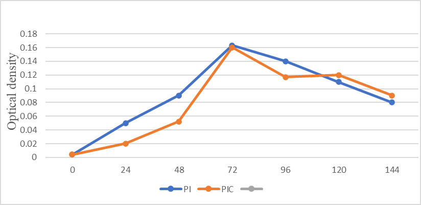
Fig. 1: Lognormal plot showing the growth of penicillium Italicum (PI) on polyethylene (PE) and penicillium Italicum control (PIC) (glucose)
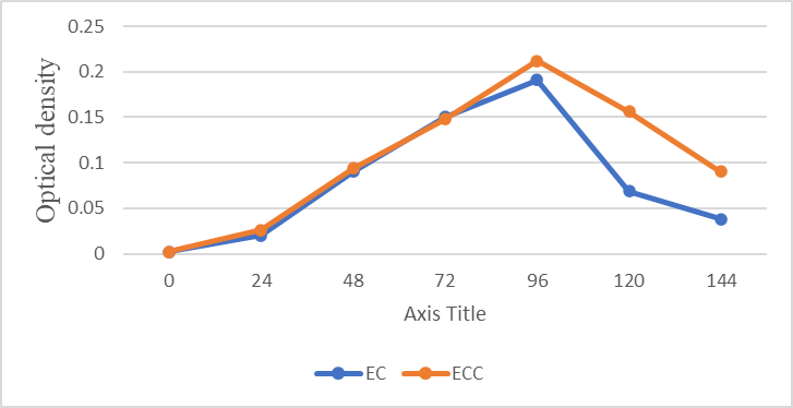
Fig. 2: Lorentzian plot showing the growth of Escherichia coli (EC) on polyethylene (PE) and Escherichia coli control (ECC) (glucose)
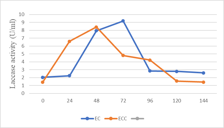
Fig. 3: Lorentzian plot showing laccase activity in Escherichia coli (EC) on polyethylene (PE) and Escherichia coli control (ECC)
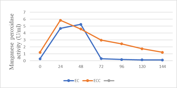
Fig. 4: Lorentzian plot showing manganese peroxidase activity in Escherichia coli (EC) on polyethylene (PE) and Escherichia coli control (ECC)
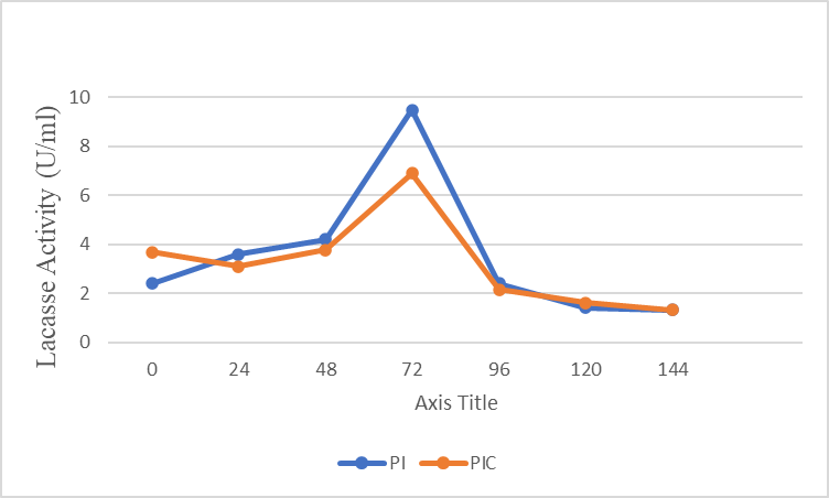
Fig. 5: Lorentzian plot showing laccase activity in penicillium italicum (PI) on polyethylene (PE) and penicillium italicum control (PIC)
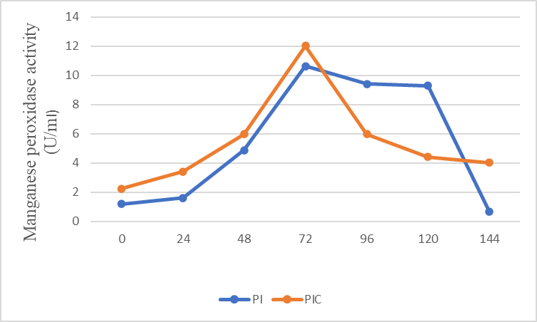
Fig. 6: Lorentzian plot showing manganese peroxidase activity in penicillium italicum (PI) on polyethylene (PE) and penicillium italicum control (PIC)
DISCUSSION
The growth rate of the microorganisms was very slow in the first two to three days of inoculation. This indicates that the organisms were not well adapted to the available carbon source. From 3rd to 5th d for fungi and bacteria, the growth started to speed up, implying that microorganisms were now well adapted to the available polyethylene (PE) carbon source. They metabolized the available carbon source for a week, after which the growth rate started declining. The activity of laccase was determined according to a modified method of [15]. This was done by monitoring the change in absorbance spectrophotometrically at 420 nm (A420), related to the rate of oxidation of 1 nm 2,2’azino-bis-(3~ethyl benzothiazoline-6-sulphonic acid) (ABTS) in 1 nm Tris-HCl buffer at pH 7.0. The enzymatic activity of laccase for penicillium Italicum (PI) and penicillium Italicum control (PIC) was monitored using optical density for 7days. The activity of the enzyme was best on day 4 for both penicillium Italicum (PI) and penicillium Italicum control (PIC), with penicillium Italicum (PI) having 9.5 and penicillium Italicum control (PIC) having 0.91. A graph for penicillium Italicum (PI)and penicillium Italicum control (PIC) was plotted and it gave the shape of a Lorentzian as shown in fig. 8.
A modified method of [16] was used to monitor manganese peroxidase (Mnp) activity over the entire biodegradation period via the oxidation of 0.24 mmol 2,2’azino-bis-(3~ethyl benzothiazoline-6-sulphonic acid) (ABTS) buffered with 50 mmol sodium acetate buffer (pH 5) in the presence of 5 mmol H2O2 at 414 nm for 5 min in a visible spectrophotometer. The enzyme activity of manganese peroxidase for penicillium Italicum (PI) was also determined using optical density for 7 d. The activity of the enzyme was best on day 4, with penicillium Italicum (PI) having 10.643 and penicillium Italicum control (PIC) having 12.033. The graph plotted for penicillium Italicum (PI) and penicillium Italicum control (PIC) gave the Lorentzian as shown in fig. 9. The enzyme activity of laccase for Escherichia coli (EC) and Escherichia coli control (ECC) was determined using optical density for a maximum of 7 d. The activity of the enzyme was at its best on day 4 for Escherichia coli (EC) with a value of 9.2, and For Escherichia coli control (ECC) on day 3, having a value of 8.42. The graph plotted for Escherichia coli (EC) and Escherichia coli control (ECC) gave the Lorentzian shape as shown in fig. 6. The enzyme activity of manganese peroxidase for Escherichia coli (EC) and Escherichia coli control (ECC) was determined using optical density. Spectroscopic methods are useful in the quantitative analysis of test samples [17]. The enzyme activity was observed to be best on day 3 for Escherichia coli (EC), having a value of 5.25, and on day 2 for Escherichia coli control (ECC) with a value of 5.83. The graph plotted for Escherichia coli (EC) and Escherichia coli control (ECC) gave the Lognormal shape as shown in fig. 7. From the results obtained, penicillium Italicum (PI) showed more activity for the enzyme manganese peroxidase when compared to laccase. While Echerichia coli (EC) showed more enzyme activity for laccase when compared to manganese peroxidase (Mp). The result of this test supports the work by [18], they observe that the presence of laccase and manganese peroxidase can aid in the degradation of polyethylene.
CONCLUSION
Escherichia coli (EC) and penicillium italicum (PI) showed Manganese peroxidase and Lacasse activities as they utilized polyethylene as a carbon source. Hence, they should be explored for biodegradation of polyethylene. We recommend that, Escherichia coli (EC) and Penicillium italicum (PI) should be used for polyethylene degradation. Further work should be done to unveil the potential of these microbial strains in biodegradation of polyethylene. Characterization of the screened enzymes should be done to optimize their production and effectiveness on plastic (polyethylene) degradation.
ACKNOWLEDGEMENT
The authors sincerely appreciate the head of the department and staffs of physical and chemical science, faculty of Basic and Applied Science, Elizade University Ilara-mokin, Ondo state, Nigeria.
FUNDING
Nil
AUTHORS CONTRIBUTIONS
Timinibefi D. Zige: laboratory demonstration, data analysis, manuscript draft; Ogunjemite O. E, laboratory demonstration supervision, manuscript review, and approval.
CONFLICT OF INTERESTS
The authors declare they have no conflict of interest.
REFERENCES
Demirbas A. Biodegradable plastics from renewable resources. Energy Sources Part A: Recovery Utilization and Environmental Effects. 2007;29(5):419-24. doi: 10.1080/009083190965820.
Andrady AL, Neal MA. Applications and societal benefits of plastics. Philos Trans R Soc Lond B Biol Sci. 2009;364(1526):1977-84. doi: 10.1098/rstb.2008.0304, PMID 19528050.
Tokiwa Y, Ugwu CU. Biotechnological production of (R)-3-hydroxybutyric acid monomer. J Biotechnol. 2007;132(3):264-72. doi: 10.1016/j.jbiotec.2007.03.015, PMID 17543411.
Siddiqui MN, Gondal MA, Redhwi HH. Identification of different type of polymers in plastics waste. J Environ Sci Health A Tox Hazard Subst Environ Eng. 2008;43(11):1303-10. doi: 10.1080/10934520802177946, PMID 18642154.
Tokiwa Y, Calabia BP, Ugwu CU, Aiba S. Biodegradability of plastics. Int J Mol Sci. 2009;10(9):3722-42. doi: 10.3390/ijms10093722, PMID 19865515.
Restrepo Florez JM, Bassi A, Thompson MR. Microbial degradation and deterioration of polyethylene-a review. Int Biodeterior Biodegrad. 2014;88:83-90. doi: 10.1016/j.ibiod.2013.12.014.
Watanabe M, Kawai F, Shibata M, Yokoyama S, Sudate Y. Computational method for analysis of polyethylene biodegradation. J Comput Appl Math. 2003;161(1):133-44. doi: 10.1016/S0377-0427(03)00551-X.
Yang J, Yang Y, Wu WM, Zhao J, Jiang L. Evidence of polyethylene biodegradation by bacterial strains from the guts of plastic-eating waxworms. Environ Sci Technol. 2014;48(23):13776-84. doi: 10.1021/es504038a, PMID 25384056.
Bombelli P, Howe CJ, Bertocchini F. Polyethylene bio-degradation by caterpillars of the wax moth Galleria mellonella. Curr Biol. 2017;27(8):R292-3. doi: 10.1016/j.cub.2017.02.060, PMID 28441558.
Pasieczna Patkowska S, Lesiuk A. Chemik. 2013;67:863-72.
Sterfan B, Czeslaw S, Zofia Z, Helena S. Biodegradation of films of polyethylene modified with starch. Studies on changes in the supramolecular structure of polyethylene. Polimery (Warsaw, Poland). 2004;49(6):424-31.
Mohan KS, Srivastava T. J Biochem Technol. 2010;2:210-5.
Cohen R, Persky L, Hadar Y. Biotechnological applications and potential of wood-degrading mushrooms of the genus Pleurotus. Appl Microbiol Biotechnol. 2002;58(5):582-94. doi: 10.1007/s00253-002-0930-y, PMID 11956739.
Wang X, Yao B, Su X. Linking enzymatic oxidative degradation of lignin to organics detoxification. Int J Mol Sci. 2018;19(11):3373. doi: 10.3390/ijms19113373, PMID 30373305.
Bourbonnais R, Paice MG, Reid ID, Lanthier P, Yaguchi M. Lignin oxidation by laccase isozymes from Trametes versicolor and role of the mediator 2,2’-azinobis(3-ethylbenzthiazoline-6-sulfonate) in kraft lignin depolymerization. Appl Environ Microbiol. 1995;61(5):1876-80. doi: 10.1128/aem.61.5.1876-1880.1995, PMID 7646025.
Hunter Christie L, Maurus R, Marcia R, Lee MH, Emma L, Raven. Harry Tong, Nham Nguyen, Michael Smith, Gary D, Brayer A, Grant Mauk. Introduction and characterization of a functionally linked metal ion binding site at the exposed heme edge of myoglobin. Proceedings of the National Academy of Sciences. 2003;100(7):3647-52. https://doi.org/10.1073/pnas.0636702100.
Vaikosen EN, Bunu SJ, Dode E, Efidi RB. Spectrophotometric fingerprinting and chemical determination of streptomycin, amikacin, neomycin, and gentamycin sulphate by condensing with Ninhydrin reagent. Int J Chem Res. 2023;7:5-10. doi: 10.22159/ijcr.2023v7i3.221.
Sowmya HV, Ramalingappa KM, Krishnappa M, Thippeswamy B. Degradation of polyethylene by Penicillium simplicissimum isolated from local dumpsite of shivamogga district. Environ Dev Sustain. 2015;17(4):731-45. doi: 10.1007/s10668-014-9571-4.